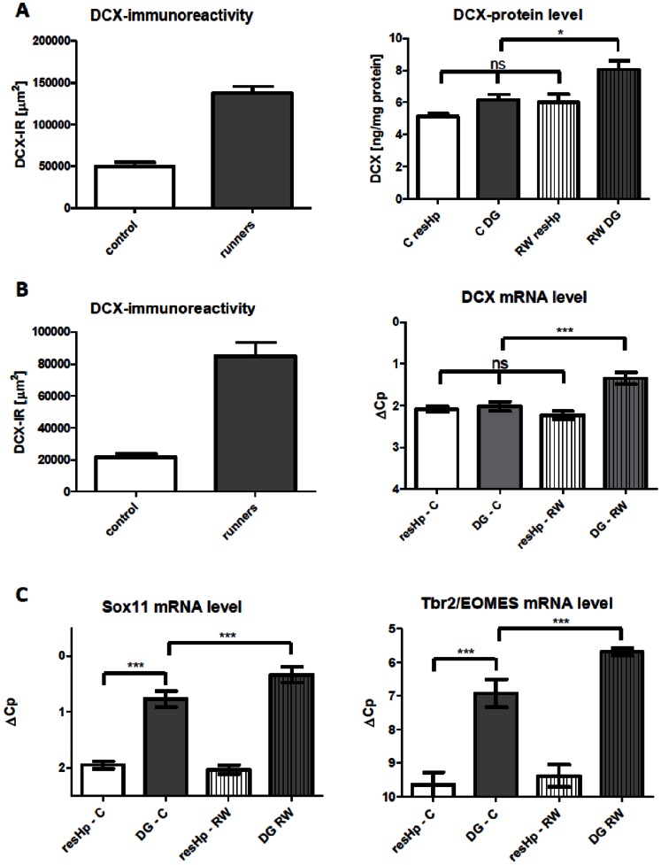
Figure 4. 14-day voluntary running wheel experiment.
In two separate experiments, adult mice with or without access to a running wheel were sacrificed after 2 weeks. The right hemisphere was dissected for either protein or mRNA-analysis while the left hemisphere was used to confirm exercise-induced increase in Dcx-IR via immunohistochemistry. A, Dcx-IR was quantified by calculating the total Dcx-IR area in µm2 for four different sections within the dorsal hippocampus (left). Bar graph of hippocampal Dcx-protein-levels in DG and resHp (right). N = 12/group. B, Dcx-IR was quantified by calculating the total Dcx-IR area in µm2 for four different sections within the dorsal hippocampus (left). Bar graph of hippocampal Dcx-mRNA-level in DG and resHp (right). N = 15/group. C, Bar graph mRNA-level in DG and resHp for Sox11 and Tbr2/EOMES. N = 15/group. Bonferroni’s Multiple Comparisons Test.

