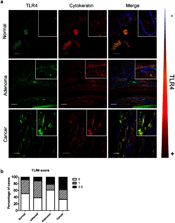
Figure 1. TLR4 is over-expressed by IECs in sporadic colorectal cancer.
a) Microarrays of human colonic tissues were stained for TLR4 (green), pan-cytokeratin (red) to detect the epithelial compartment, and counterstained with DAPI (blue). Normal tissue, adenoma and cancer. Epithelial TLR4 is best visible in the merged image (yellow) Scale bar: 100 µm. White squares highlight the areas that were magnified. b) Graph showing level of TLR4 expression in the colonic epithelium increases from normal to adenoma and cancer in human tissue microarrays. Data represent percentage of cases with TLR4 staining in IECs with score 0 (negative), 1 (low positive), and 2–3 (medium and high positive), for each histopathologic category: normal (n = 12), inflamed (n = 8), adenoma (n = 23), and cancer (n = 52).

