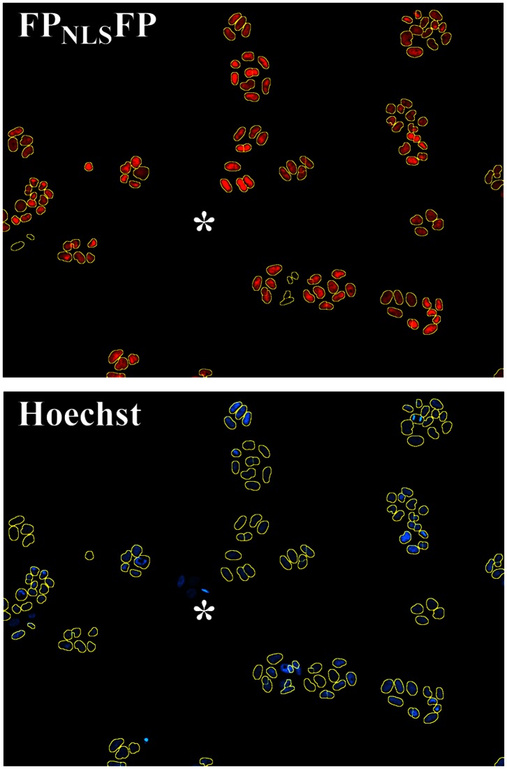
Figure 2. Segmentation of cell nuclei marked with FPNLSFP.
The boundaries of objects with contiguous FPNLSFP expression established by a commercial analysis software (yellow circles) marked the boundaries of nuclei stained with the DNA binding dye Hoechst 33342. *, colony of cells in which FPNLSFP expression is lost sporadically.

