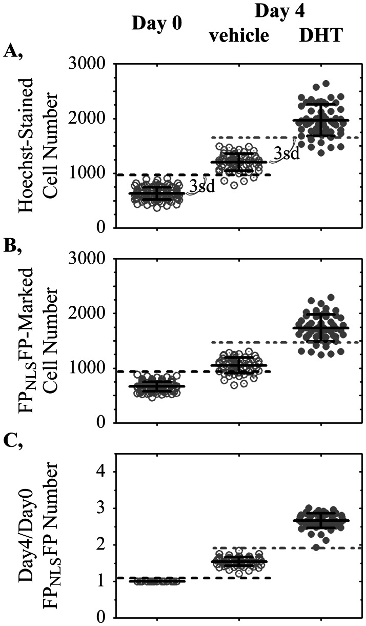
Figure 3. Improved well-to-well reproducibility in cell growth measurement enabled by the FPNLSFP live cell nuclear marker.
Variations in cell numbers plated in each well (Day 0) obscured the ability to reliably detect an increase in cell number after four days of slow growth by LNCaP-C4-2 prostate cancer cells treated with vehicle or 0.2 nM DHT. Each symbol represents the numbers of A, Hoechst 33342-stained nuclei or B, FPNLSFP-marked nuclei segmented in each well. C, Dividing the number of FPNLSFP-marked cells on Day 4 by the baseline (Day 0) number of FPNLSFP-marked cells in the same well improved the reproducibility of growth measurement. Dotted lines, three standard deviations (3sd) above the mean Day 0 (black dotted line) or Day 4 vehicle-treated (gray dotted line) measurements are shown. The 3sd cut-offs were used to determine the number of, respectively, vehicle-treated and DHT-treated wells that were scored falsely in the Day 0 and vehicle-treated wells.

