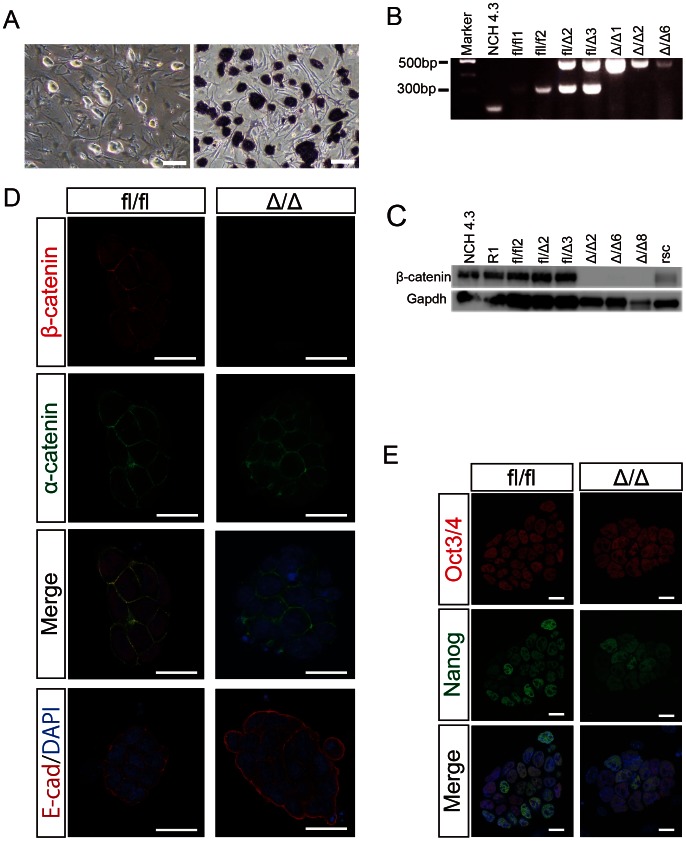
Figure 1. Characterization of embryo-derived β-catΔ/Δ mESCs.
(A): Morphological appearance of β-catΔ/Δ mESCs is shown in the left panel. ALP staining of β-catΔ/Δ mESCs is shown in the right panel. Scale bars are 150 µm. (B): Electrophoretic analysis of the genotyping PCR for wild-type (NCH4.3), β-catfl/fl (fl/fl1 and fl/fl2), β-catfl/Δ (fl/Δ2 and fl/Δ3) and β-catΔ/Δ (Δ/Δ1, Δ/Δ2 and Δ/Δ6) mESCs. (C): Western blots of β-catenin and Gapdh in wild-type (NCH4.3 and R1), β-catfl/fl (fl/fl2), β-catfl/Δ (fl/Δ2 and fl/Δ3), β-catΔ/Δ (Δ/Δ2, Δ/Δ6 and Δ/Δ8) and β-catenin rescued β-catΔ/Δ mESCs (rsc). (D and E): Immunofluorescence staining for β-catenin (red), α-catenin (green), E-cadherin (red) and Oct3/4 (red), Nanog (green) of β-catfl/fl and β-catΔ/Δ mESC colonies as observed under confocal microscopy. Nuclei are stained for DAPI (blue). Scale bars in (D) and (E) are 20 µm.

