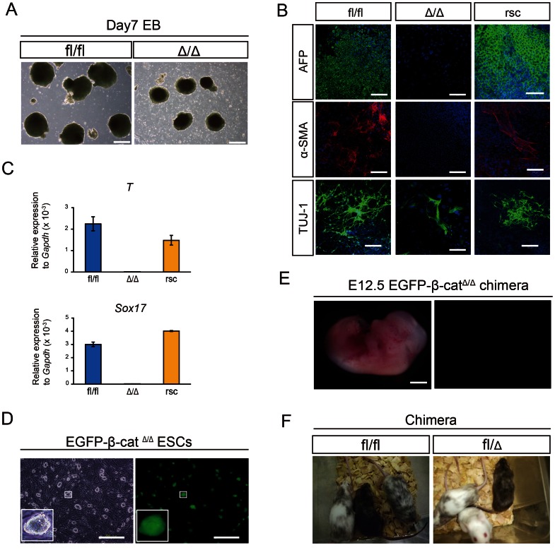
Figure 2. Differentiation potential of embryo-derived β-catΔ/Δ mESCs.
(A): Phase contrast pictures of EBs derived from β-catfl/fl and β-catΔ/Δ mESCs on day 7 of differentiation. Scale bars are 500 µm. (B): Immunofluorescence staining for Afp (green), α-SMA (red) and Tuj-1 (green) of β-catfl/fl, β-catΔ/Δ and β-catenin rescued β-catΔ/Δ mESCs on day 14 of differentiation. Nuclei are stained for DAPI (blue). Scale bars are 200 µm. (C): Expression levels of Sox17 and Brachyury(T) relative to Gapdh in β-catfl/fl (blue bar), β-catΔ/Δ (light blue bar) and β-catenin rescued β-catΔ/Δ (yellow bar) mESCs on day 6 of differentiation. (D): Fluorescence microscopic images of EGFP-β-catΔ/Δ mESC colonies. They constitutively expressed EGFP. Scale bars are 500 µm. (E): Fluorescent stereomicroscopic image of an embryo on E12.5 generated from blastocysts injected with EGFP-β-catΔ/Δ mESCs. The contribution of β-catΔ/Δ mESCs were barely detected anywhere in the whole body. Scale bars are 2 mm. (F): Chimeras generated by injection of β-catfl/fl and β-catfl/Δ mESCs into ICR host blastocysts. Offspring coat color demonstrates high contribution chimeras.

