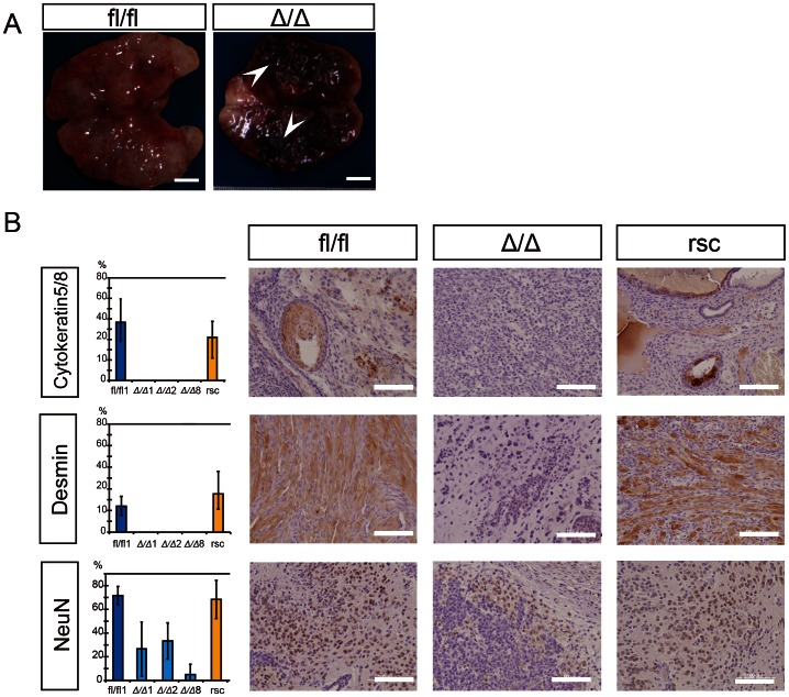
Figure 3. Immunohistochemical examination of tumors generated from ESCs either β-catΔ/Δ or β-catfl/fl.
(A): The gross image of a tumor mass derived from β-catfl/fl mESC is shown on the left and that of a tumor derived from β-catΔ/Δ mESCs is shown on the right. β-catΔ/Δ tumor mass was characterized by extensive intra-tumoral hemorrhage (white arrow head). Scale bars are 5 mm. (B): Immunohistochemical staining for Cytoketratin 5/8, Desmin and Neuronal nuclear antigen (NeuN) in tumors derived from mESCs of β-catfl/fl, β-catΔ/Δ or res-β-catΔ/Δ. Tissue sections of β-catΔ/Δ tumors displayed high level staining only for the neuronal differentiation marker NeuN, while there was no detectable staining for Cytoketratin 5/8 or Desmin. Tissue sections of both β-catfl/fl and res-β-catΔ/Δ tumors displayed multiple differentiations as shown in three markers’ positive staining. The left bar graph shows percentages of Cytoketratin 5/8, Desmin and NeuN positive areas with standard deviation (n = 3) as the vertical axis and each tumor as the horizontal axis. Scale bars are 100 µm.

