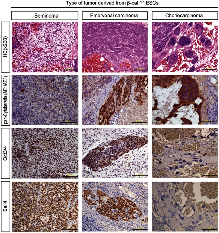
Figure 4. Histological and immunohistochemical examination of β-catΔ/Δ tumors.
Careful examination of numerous tumor sections stained with hematoxylin and eosin (HE) from different β-catΔ/Δ mESC lines generated independently in separate animals revealed that each β-catΔ/Δ tumor contained three subtypes (seminoma, embryonal carcinoma, choriocarcinoma) of carcinomatous germ cell tumor components. The “seminoma” component is characterized by large uniform dispersed tumor cells displaying clear cytoplasm and distinct cell membrane. Histologic features of the “seminoma” were “fried-egg” appearance: central nucleus with nucleolus, clear cytoplasm and well-defined cell borders. The “embryonal carcinoma (EC)” displayed a papillary pattern, with cohesive clustered growth and marked cytologic atypia. The “choriocarcinoma” was identified by syncytiotrophoblastic giant cells with extensive hemorrhage. The syncytiotrophoblastic giant cells had huge, pleomorphic nuclei and abundant eosinophilic cytoplasm. The “seminoma” displayed very high levels of nuclear OCT4 and Sall4 immunoreactivity, but was negative for pan-cytokeratin. “EC” was strongly positive for pan-cytokeratin, Oct3/4 and Sall4. The syncytiotrophoblastic giant cells of the “choriocarcinoma” showed positive cytoplasmic staining for pan-cytokeratin, but negative nuclear immunostaining for Oct3/4 and Sall4. Scale bars are 100 µm.

