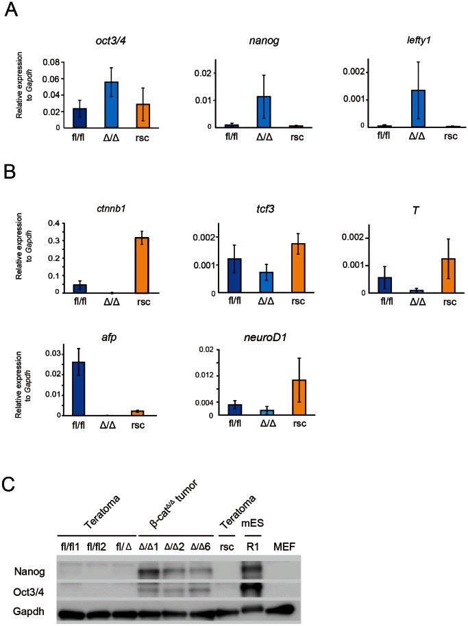
Figure 6. Quantitative PCR and western blot examination of wild-type ESCs derived teratomas, res-β-catΔ/Δ teratomas and β-catΔ/Δ tumors.
(A and B): Expression levels of self-renewal marker genes (oct3/4, nanog, lefty1) and the downstream genes of Wnt/β-catenin signaling (Ctnnb1, tcf3, T) and early differentiation markers (afp, neuroD1, T) relative to Gapdh in β-catfl/fl (blue bar), res-β-catΔ/Δ (yellow bar) and β-catΔ/Δ (light blue bar) mESC derived teratomas or tumors. (C) Western blots of Nanog, Oct3/4 and Gapdh for teratomas derived from β-catfl/fl (fl/fl1,fl/fl2), β-catfl/Δ, res-β-catΔ/Δ (rsc) and R1 mESCs, tumors derived from β-catΔ/Δ (Δ/Δ1, Δ/Δ2, Δ/Δ6) mESC. MEF was used as a control. Abbreviation: MEF, mouse embryonic fibroblast.

