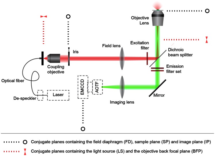
Figure 1. Diagram of the custom-modified epi-luminescence imaging system employed for single-UCNP and spectral imaging.
A wide-field inverted epi-luminescence microscope was modified to allow external fiber-coupled laser illumination. The optical fiber was dithered to average out speckles. The excitation light was configured to uniformly illuminate the field-of-view at the sample plane via a modified Köhler illumination scheme. The sample plane was imaged using an EMCCD camera, optionally mounted with an AOTF for hyperspectral imaging. An adjustable iris diaphragm allowed reduction of the field-of-view to restrict imaging to several single UCNP particles and small clusters.

