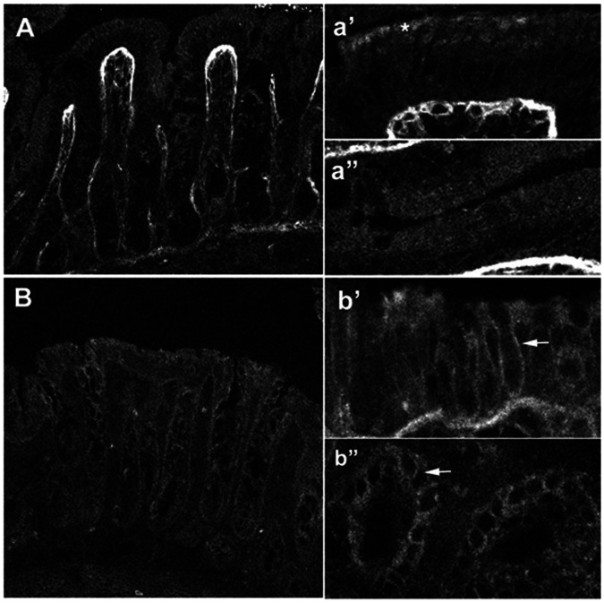
Figure 4. Immunocytochemical localization of NBCn1-like proteins in rat proximal and distal colon.
[A, a’, a”] Rat proximal colon demonstrates weak epithelial staining for NBCn1 along the crypt to surface axis. The connective tissue underlying the epithelial cells is strongly fluorescent. Higher magnification of the surface cells [a’] shows a rather punctuate apical signal [*]. High magnification of epithelial cells in the crypt region (a”) demonstrates a weak diffuse localization. [B, b’, b”] Rat distal colon shows stronger staining for NBCn1. High magnifications of surface cells [b’] and crypt cells [b”] clearly demonstrate basolateral membrane localization [arrows]. Smooth muscle cells were also strongly stained (data not shown).

