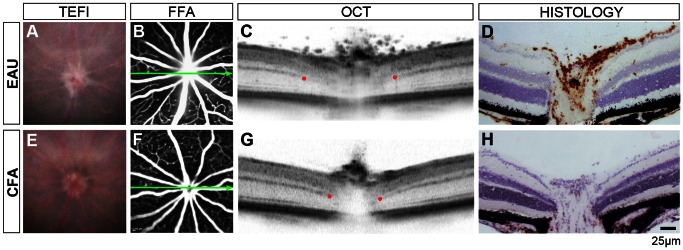
Figure 1. EAU in the C57BL/6J mouse induces optic nerve head changes detectable on OCT.

Representative images from an eye 15 days post-induction of EAU (A–D) and matched CFA control (E–H) using four modalities. Blurred optic disc margins on TEFI (A) and disc hyperfluorescence on FFA (B) are seen in EAU. Green arrows show the location and orientation of the OCT scan. Red dots indicate where the discrete signal from the outer plexiform layer becomes obscured by disc enlargement, as a landmark to compare between eyes. Note a wider separation and vitreous opacities in EAU (C). Matched histology demonstrates CD45+ infiltration (DAB in brown with haematoxylin counterstain) during EAU (D). With TEFI alone (E), it is possible that changes in disc appearance would erroneously be identified as disease with infiltration in the CFA control, as OCT and immunohistochemistry do not corroborate any evidence of disease.
