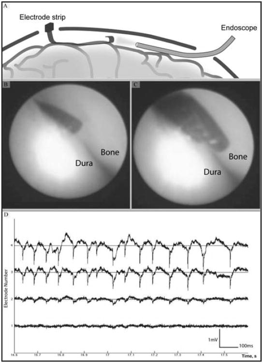
Figure 4.
Long strip electrode suitable for acute recording. A) A representation of a strip electrode inserted through a very small (10 mm) burr hole simultaneously with an endoscope for real time imaging of electrode position. B) and C) Real time images of electrodes in contact with the dura, taken from the scope which was situated on the opposite side of the location of electrode entry. D) Epileptic activity observed from an acute recording using the same long strip electrode array. Spontaneous epileptic activity recorded under anethesia from 4 neighboring electrode sites shows the signal amplitude of up to 2 mV, droping over a spatial scale resolvable only by micro-ECoG arrays.

