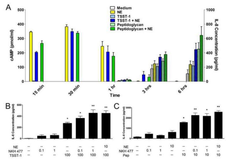
Figure 5.
Norepinephrine (NE)-based enhancement of IL-8 responses in HVECs is dependent on cAMP signaling. A) HVEC-2 cells were incubated with toxic shock syndrome toxin-1 (TSST-1, 100 μg/ml), peptidoglycan (10 μg/ml), or NE (10 μM) over a total period of 6 hours. At various time points, supernates were collected to determine IL-8 concentrations secreted into the medium and cells were lysed to look at intracellular levels of cAMP. Plain bars indicate cAMP levels, while hatched bars indicate IL-8 levels. N=6, two independent experiments. Similar results were obtained with HVEC-1 cells. B) HVEC-2 cells were incubated with TSST-1 (100 μg/ml), NE (10 μM), or the forskolin analog NKH 477 (1 μM). **Significantly higher than all other conditions and *significantly higher than medium only and both NKH 477 only controls by one-way ANOVA [F(6,35)=32.66, p<0.0001] and Tukey’s post-hoc test. N=6, two independent experiments. C) HVEC-1 cells were incubated with peptidoglycan (10 μg/ml), NE (10 μM), or NKH 477 (1 μM). **Significantly higher than all other conditions and *significantly higher than medium only, NE only, and both NKH 477 only controls by one-way ANOVA [F(7,34)=43.58, p<0.0001] and Tukey’s post-hoc test. N=6, two independent experiments. Error bars represent the standard error of the mean.

