
Fig. 4.
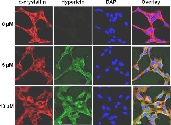
Colocalization of hypericin with α-crystallin in unirradiated HLE cells. HLE cells were incubated with either DMSO or hypericin in DMSO for 1 h at 37 °C in the dark. Following washing and fixation, cells were stained with a mixture of mouse monoclonal anti-αA and anti-αB crystallin (red) followed by staining with Alexa Fluor anti-mouse 633 and staining with DAPI to visualize cell nuclei (blue). Natural hypericin fluorescence (ex 561, shown in green) was used to image the photosensitizer presence in the cells. Alpha-crystallin (red), hypericin (green), DAPI (blue), and overlay of all three.