
Fig. 6.
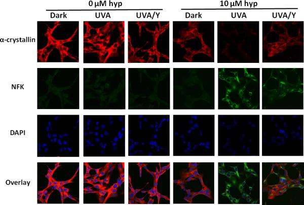
Partial protection of UVA-irradiated, hypericin-containing HLE cells by blocking of wavelengths <400 nm: The effect on NFK and α-crystallin detection. HLE cells were incubated with either DMSO (0 μM) or hypericin in DMSO (10 μM) for 1 h at 37 °C in the dark. After washing with buffer, the cells were either kept in the dark, exposed to 40 min of UVA irradiation, or exposed to 40 min of UVA irradiation through a yellow filter that removed wavelengths < 400 nm (UVA/Y). Fixed cells were stained simultaneously with a mixture of mouse monoclonal anti-αA and anti-αB-crystallin and rabbit polyclonal anti-NFK followed by staining simultaneously with Alexa Fluor anti-mouse 633 and Alexa Fluor anti-rabbit 488. DAPI was used to stain cell nuclei. Alpha-crystallin (red), NFK (green), DAPI (blue) and overlay of all three.