
Fig. 8.
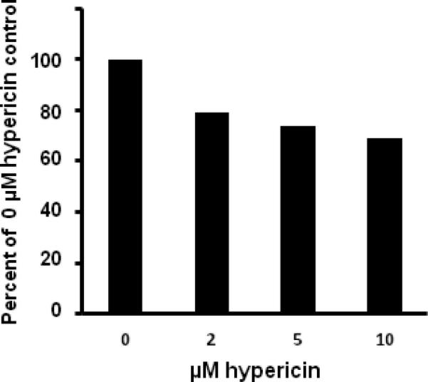
Cell viability after treatment with hypericin and UVA irradiation. Cells seeded into the wells of microtiter plates and allowed to grow until 80–90% confluence were incubated with either DMSO (0 μM) or hypericin in DMSO for 1 h at 37 °C in the dark. After washing with buffer the cells were exposed to 40 min of UVA irradiation and then assayed for viability. The data presented represent the average for 3 wells per treatment and are expressed as a percentage of the positive (0 μM hypericin) control.