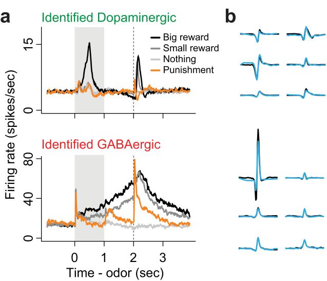
Figure 3. Electrophysiological properties of optogenetically identified dopamine and GABA neurons recorded in vivo.
Electrophysiological recordings were performed in vivo in two strains of transgenic mice expressing channelrhodopsin2 in either dopamine or GABA neurons. During recording sessions, subjects were engaged in an odor-outcome association task with positive, negative and neutral outcomes. (A) Average responses of identified dopamine neurons (top; n=26) and identified GABA neurons (bottom; n=17) to odor cues (0-1 seconds, grey bar) and associated outcomes (delivered at 2 seconds, dashed line). Note that GABA neurons increase their firing rate during reward-predictive cues and this activity is sustained until the expected time of reward delivery. (B) Spontaneous (black) and optically evoked (blue) waveforms from 6 individual neurons recorded in mice expressing channelrhodopsin2 in dopamine (top) or GABA neurons (bottom). Close correspondence between spontaneously occurring and optically evoked waveform shapes was one of the criteria used to identify the neurotransmitter content of recorded neurons. Adapted with permission from Cohen et al. (2012).

