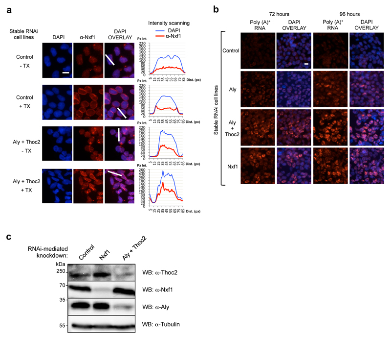
Figure 7. Thoc2 plays a role in Nxf1 localisation and mRNA export.
(a) Nuclear envelope association of Nxf1 is impaired in vivo when both TREX components Aly and Thoc2 are depleted by RNAi. Localisation of Nxf1 was analysed by immunostaining of Control or (Aly+Thoc2) knockdown cell lines after 96h of induction of miRNA expression using Nxf1 antibody. Where indicated (+ TX), cells were treated with 0.5% Triton X-100 before fixing. The Y-axis of the graphs represents the pixel intensity (Px Int.) and the X-axis the distance in pixels (Dist. (px)). (b) A strong block of bulk poly(A)+ RNA export is observed in Nxf1 RNAi and (Aly+Thoc2) RNAi cell lines. Oligo (dT) FISH on stable 293 cell lines expressing miRNAs targeting the indicated genes. The time points refer to the time following induction of miRNAs expression. Cells were treated with actinomycin D for 2 hours prior to FISH to reduce nascent RNA signals. All pictures are taken at the same exposure level. The white horizontal bars in (a) and (b) represent 10 μm. (c) Western Blot analysis of the indicated cell lines. The expression of the miRNAs is under the control of a tetracycline inducible promoter. Extracts were prepared 96 hours post-induction of miRNA expression and analysed using the indicated antibodies.

