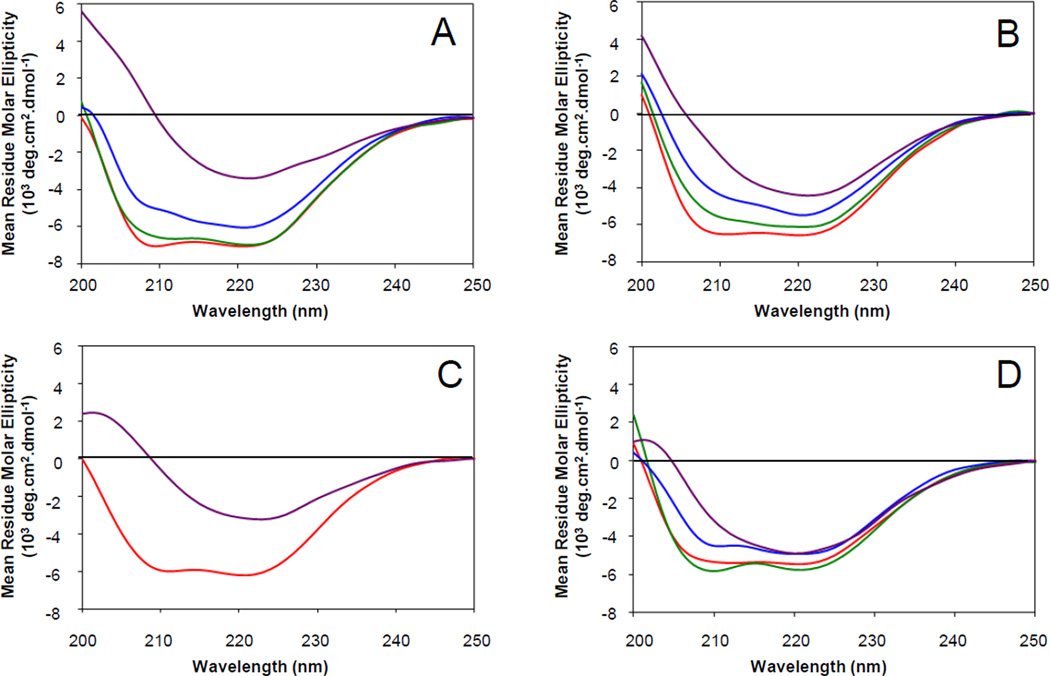
Fig. 3.
Effect of cholesterol on secondary structure of HIV gp41 fusion domain in lipid bilayers of different lipid compositions. Far-UV circular dichroism spectra of HIV gp41 fusion domain bound to small unilamellar vesicles at a peptide:lipid ratio of 1:100 were recorded at room temperature. The lipid bilayers were composed of (A) POPC:POPG (4:1), (B) POPC:POPS (4:1), (C) POPC:POPS:PI (12:2:1), or (D) POPC:POPE:SM:POPS:PI (20:5:2:2:1) with 0% (red), 10% (green), 20% (blue), 30% (A and B, purple), or 33% (C and D, purple) cholesterol.

