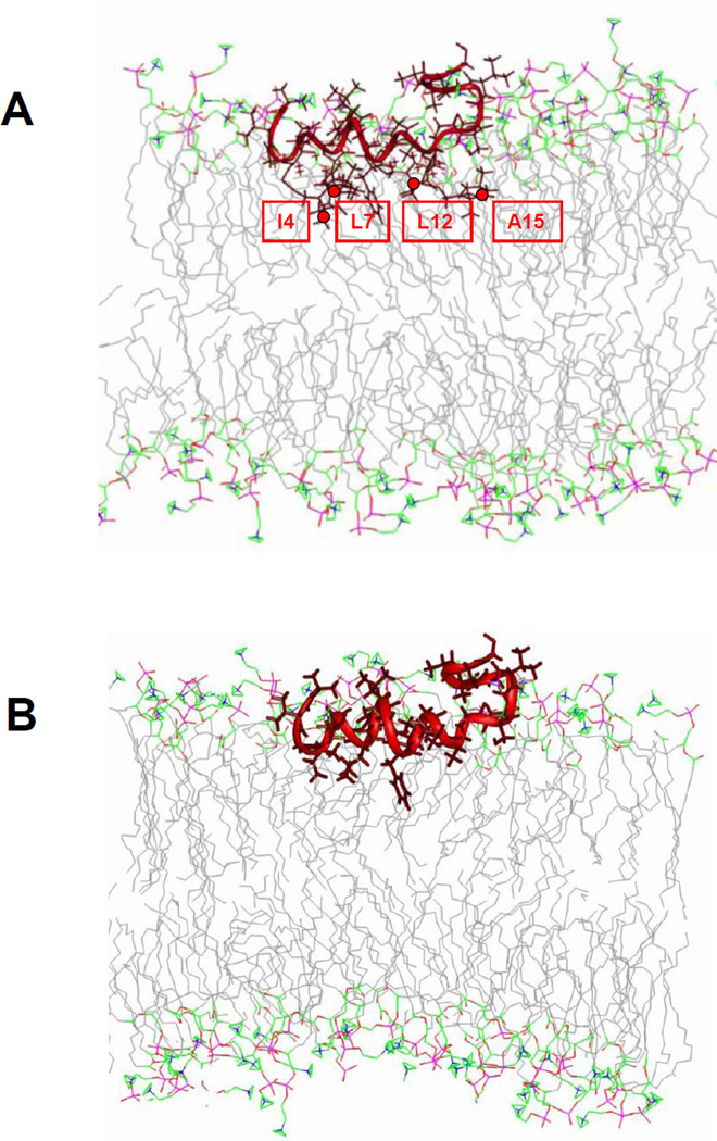
Fig. 5.
Model of the α-helical HIV gp41 fusion domain docked to a lipid bilayer based on power saturation experiments performed in POPC:POPG (4:1). (A) Docked fusion domain (PDB accession code 2PJV 11) with four cysteines substituted in positions 4, 7, 12 and 15 and with nitroxide spin labels attached in all four positions. (B). Same as in (A), but with restored native HIV gp41 fusion domain sequence. The green lines represent the average positions of lipid phosphate groups relative to the fusion domains. The positions of spin label nitroxides are marked with red circles.

