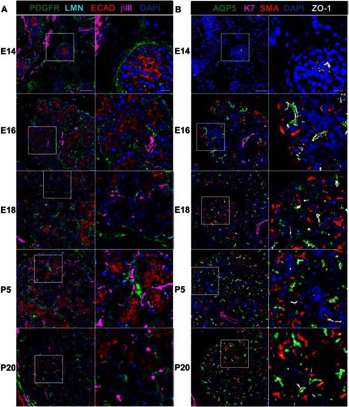
Fig. 1. MxIF analysis of mouse submandibular salivary gland morphogenesis.

MxIF of a developmental TMA including embryonic stages (E14, E16, E18) and postnatal stages (P5 and P20) was performed using sequential application of directly conjugated antibodies to detect multiple markers of tissue structures and cell types on the same tissue sections. (A) Tissue compartments. The epithelium, mesenchyme, neurons, and basement membranes was detected using antibodies directed towards E-cadherin (ECAD, red), platelet-derived growth factor (PDGFR, green), βIII tubulin (bIII, magenta), and laminin (LMN, cyan), respectively. (B) Epithelial differentiation. Maturation of the proacinar, ductal, and myoepithelial cell types was detected using antibodies to aquaporin 5 (AQP5, green), cytokeratin 7 (K7, magenta), and smooth muscle α-actin (SMA, red); maturation of the cell–cell adhesions was monitored using an antibody to zonula occludens-1 (ZO-1, white). DAPI was used to stain the nuclei in both A and B. Scale bars: 50 µm zoom level one and 10 µm zoom level two.
