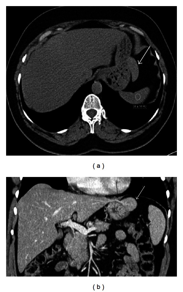
Figure 13.

Nonenhanced axial CT image (a) of a 48-year-old female patient reveals a soft tissue mass (arrow) nearby greater curvature of the stomach. On coronal reformatted contrast enhanced CT image, there is a stalk (black arrow) between left lobe of the liver and the mass (white arrow) indicating accessory liver.
