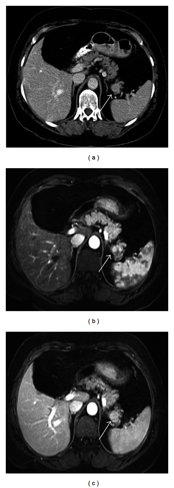
Figure 4.

Axial contrast enhanced CT image (a), contrast enhanced T1-weighted image at arterial (b) and venous phases (c) show an intrapancreatic nodular mass in a 63-year-old female patient. The mass has similar density at CT and contrast enhancement pattern at MR images with the spleen indicating an intrapancreatic accessory spleen.
