
Figure 1.
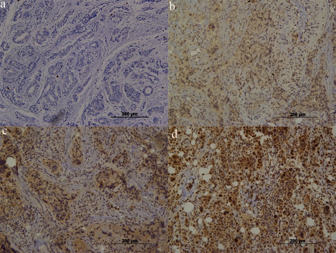
HIF1A expression in breast carcinoma. Note that homogeneous staining can be observed in all samples. We classified the samples according to staining intensity from 0 to 3+. Tumors classified as 1+ to 3+ were considered to have positive HIF1A expression. (a) Negative expression (200×); (b) weak positive (1+) expression (200×); moderate positive (2+) expression; (d) strong positive (3+) expression (200×).