
Figure 1.
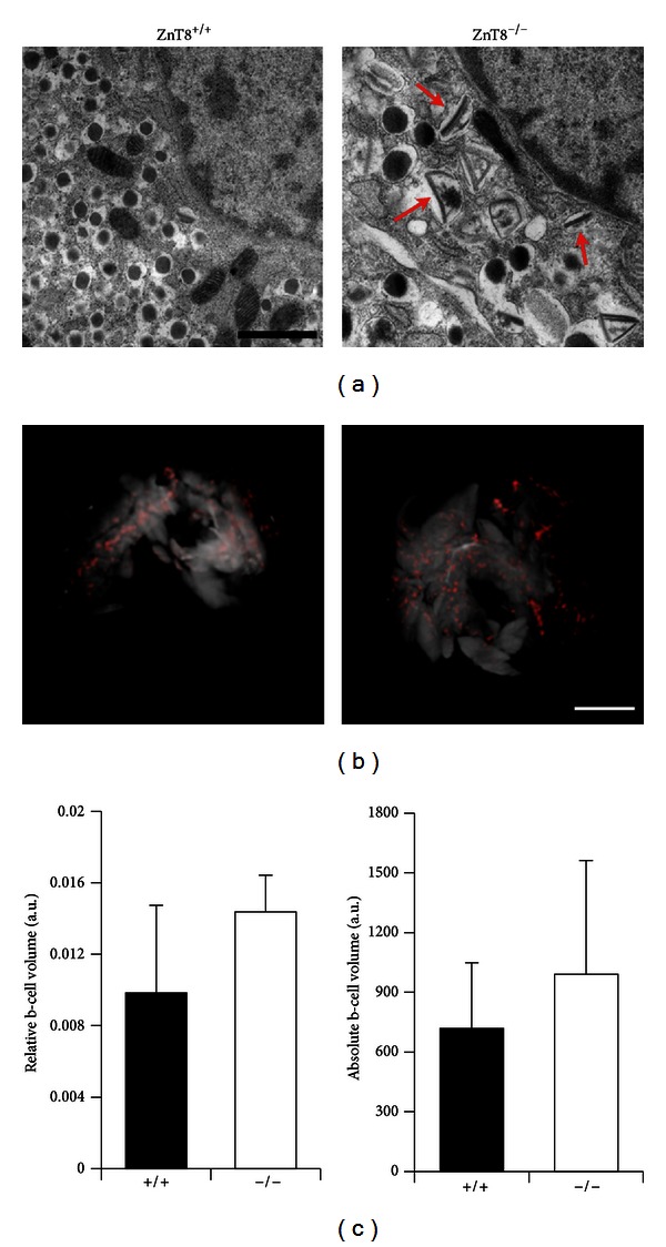
Electron Micrographs and Optical Projection Tomography (OPT) in ZnT8+/+ and ZnT8−/− mice. (a) Transmission electron microscopy images of isolated islets from ZnT8+/+ and ZnT8−/− male mice at high magnification (scale bar 1 mm) reveals the appearance of rod-shaped core granules in ZnT8−/− cells, indicated by red arrows (n = 3 mice). Sections were cut and images were acquired by Dr. Raffaella Carzaniga and Ms. Katrin Kronenberger. (b) Representative three-dimensional OPT projections of whole fixed and permeabilised pancreas from ZnT8−/− and ZnT8+/+ mice. In red are the insulin positive structures (β cells). The overall shape of the whole pancreas was visualized as autofluorescence and is apparent as white/grey shading. Scale bar = 1 cm. (c) Relative (right panel) and absolute (left panel) β-cell volume (n = 2 pancreata per genotype).