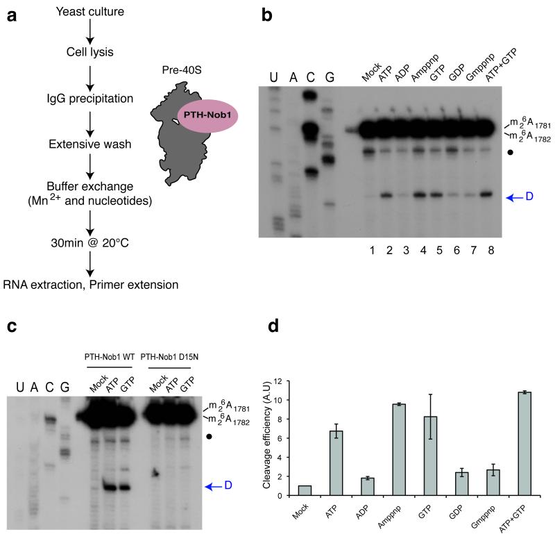
Figure 2.
In vitro cleavage in pre-40S particles is stimulated by ATP or GTP addition. (a) Diagram representing steps in the assay for in vitro 40S subunit maturation. (b) Primer extension analysis showing the activation of in vitro site D cleavage in pre-40S particles by nucleotide addition. The strong upper stops result from termination at the sites of 18S rRNA base-methylation at A1779 and A1780. These modifications precede site D cleavage in vivo. The blue arrow indicates site D. A closed circle indicates an additional primer extension stop seen in some experiments, which corresponds to a run of G-C base-pairs at the foot of H45 in the 18S rRNA. The nucleotides indicated were added at 1mM. (c) Comparison of cleavage stimulation observed with pre-40S particles purified using wild-type (PTH-Nob1 WT) or the catalytically inactive PTH-Nob1D15N protein. (d) Variation of cleavage efficiencies in relation to nucleotide addition. Signals obtained for cleavage at site D in panel (b) were quantified, corrected for RNA loading (using a common stop in the ITS1 as a standard) and normalized to the mock-treated sample (set to 1). The average of three independent experiments is shown, with the standard deviation indicated on top of the histogram.

