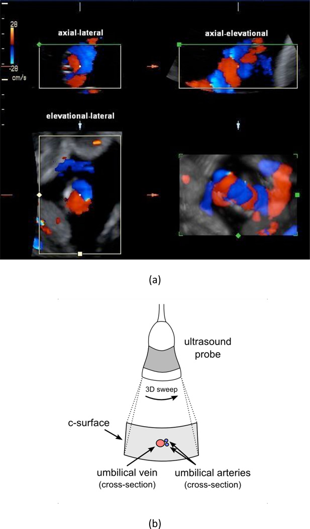
Figure 2.
(a) Four-panel view of a single 3D color flow acquisition of the umbilical cord of Patient 6a (P6a see Figure 6). The four views are as follows: upper-left is axial-lateral, upper-right is axial-elevational, bottom-left is elevational-lateral (i.e., the c-surface), and bottom-right is a rendered 3D reconstruction. The upper-left, upper-right, and bottom-left panels coincide at the center point that is marked in each view. Arteries are shown in blue and the vein is shown in red. The schematic in (b) illustrates the orientation of the probe and the corresponding c-surface in the elevational-lateral imaging plane. The vessel colors in (b) are opposite of convention in order to match the directionality in (a). The entire umbilical cord passes through the c-surface but only the cross-sections of the umbilical arteries and umbilical vein are illustrated in (b).

