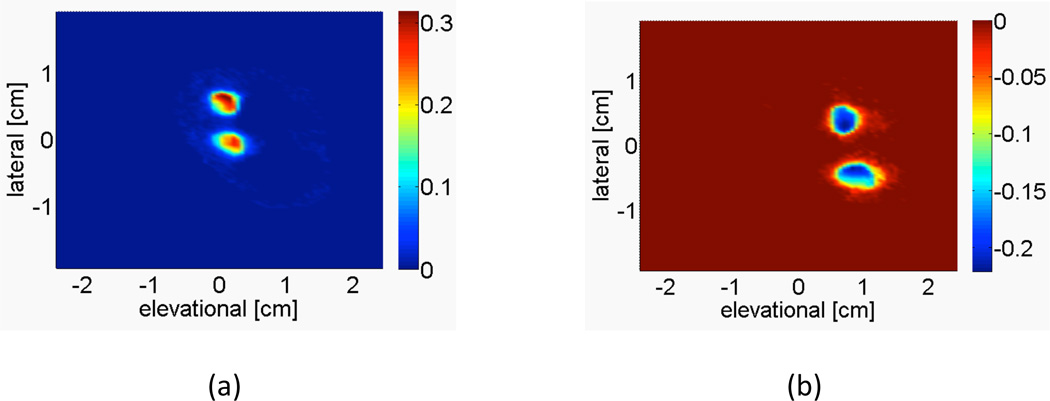
Figure 3.
Representative image from sheep subject 5 showing the power-weighted Doppler-measured velocities of the c-surface intersecting the (a) fetal umbilical arteries and (b) fetal umbilical veins, at a depth of 1.87 cm. Integrating over this c-surface yields the volume flow estimate. The color bar indicates the average power-weighted Doppler-measured velocity in m/s.

