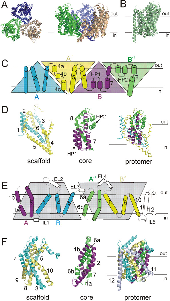
Fig. 3. Structural makeup of GltPh and LeuT.
A) GltPh assembles as a bowl-shaped trimer. Left, extracellular view. Right, view parallel to the membrane. Individual monomers are colored wheat, blue, green. B) LeuT monomer, viewed parallel to the membrane. C) Primary structure of a GltPh monomer. First inverted repeat (AA−1: blue, yellow) and second inverted repeat (BB−1: magenta, green) displayed as triangles. D) Structural relationship of internal repeat structures in GltPh. Scaffold domain, left. Core domain, middle. Protomer fold, right. TMs colored as in C. E) Primary structure of LeuT. Inverted repeat defined by gray shaded area. TMs 1–2 (A: magenta) and 6–7 (A−1: green), as well as TMs 3–5 (B: blue) and 8–10 (B−1: yellow), are symmetrically related. F) Structural relationship of internal repeat structures in LeuT. Scaffold domain, left. Core domain, middle. Protomer fold, right. TMs colored as in E. TMs 11 and 12 shown only in the protomer fold for clarity.

