
Figure 4.
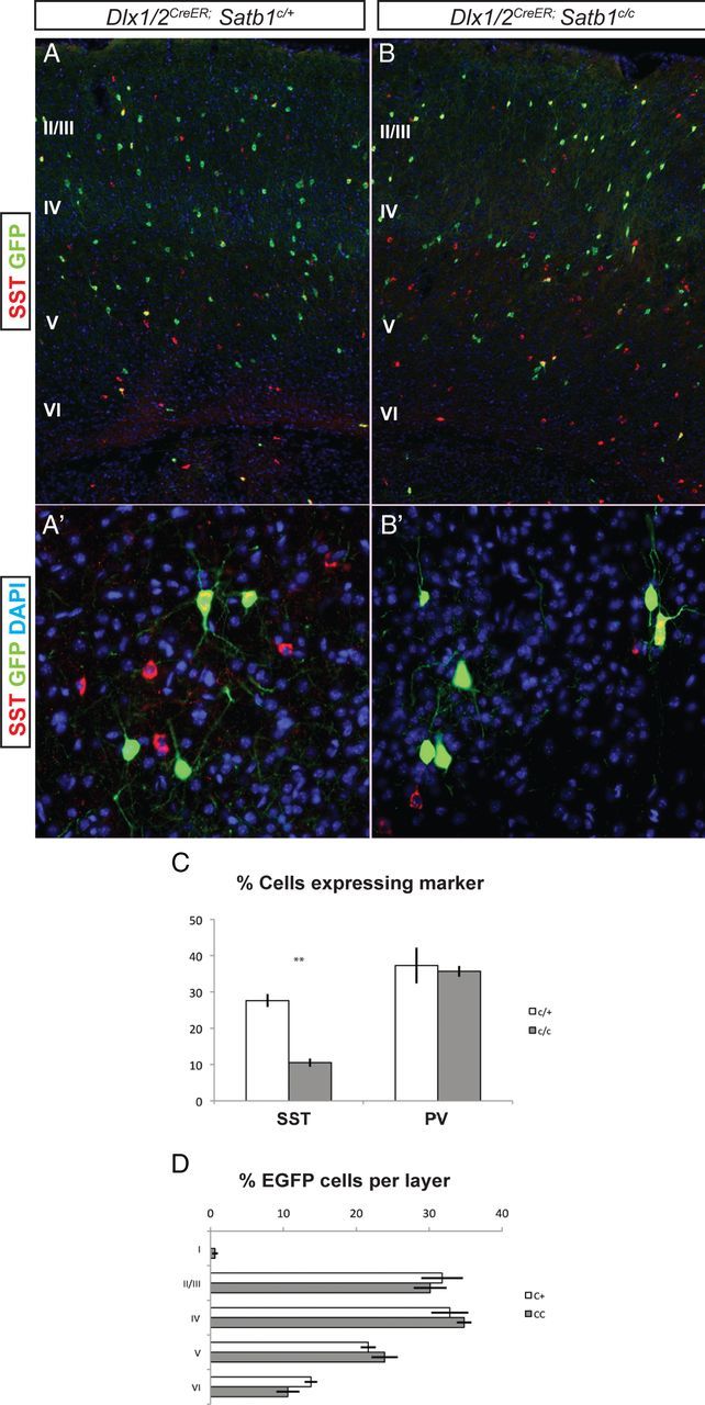
SST INs require postnatal expression of Satb1. Tamoxifen was administered to Dlx1/2CreER;Satb1 conditional animals on P1, and animals were killed at P21 for analysis. All analysis was done on animals positive for the RCE:LoxP reporter allele for Cre-mediated EGFP expression. A, B, The numbers of EGFP-positive INs were similar between Dlx1/2CreER;Satb1c/+ controls (A) and Satb1c/c animals (B). A', B' Higher-powered view of deep-layer cells positive for EGFP after P1 tamoxifen administration. C, Quantification of the percentage of EGFP cells expressing MGE markers. In Dlx1/2CreER;Satb1c/+ animals, 27.6 ± 1.7% of cells were SST-positive, compared with 10.5 ± 1.1% in Satb1c/c animals following P1 administration of tamoxifen. D, The number of cells destined for each cortical layer was similar between Satb1c/+ and Satb1c/c animals. n = 4 for Satb1c/+ animals and n = 5 for Satb1c/c animals. **p < 0.01.