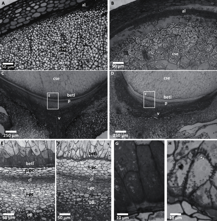
Fig. 4.
Cellular defects in pgd3-umu1 kernels. Toluidine blue-stained semithin sections of 16-DAP kernels of wild-type sibling (A, C, E, G) and pgd3 (B, D, F, H). (A, B) Apical region of the seed showing central starchy endosperm (cse) and aleurone (al); bars = 50 µm. (C, D) Basal region showing the cse, basal endosperm transfer layer (betl), pedicel (p), and vascular tissue (v); boxes indicate regions magnified in E and F; bars = 250 µm. (E, F) Pedicel region showing the betl, nucellar placento-chalazal layer (n-pc), closing layer (cl), integumental placento-chalazal layer (i-pc), and pedicel parenchyma (pp); bars = 50 µm. (G, H) Basal endosperm transfer layer cells; white arrows indicate short secondary cell-wall ingrowths found in pgd3 mutants; bars = 10 µm.

