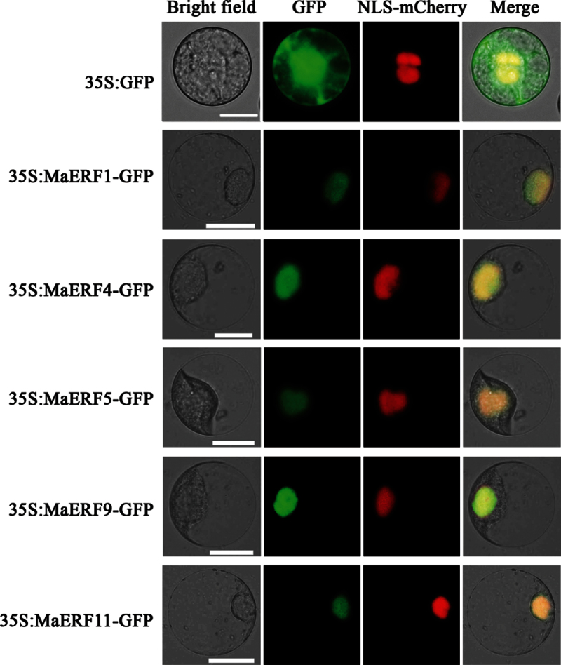
Fig. 1.
Subcellular localization of MaERFs in tobacco BY-2 protoplasts. Protoplasts were transiently transformed with MaERF–GFP constructs or GFP vector using a modified PEG method. GFP fluorescence was observed with a fluorescence microscope. VirD2NLS-mCherry was included in each transfection to serve as a control for successful transfection, as well as for nuclear localization. Images were taken in a dark field for green fluorescence, while the outline of the cell and the merged were photographed in a bright field. Bars, 25 μm (this figure is available in colour at JXB online).

