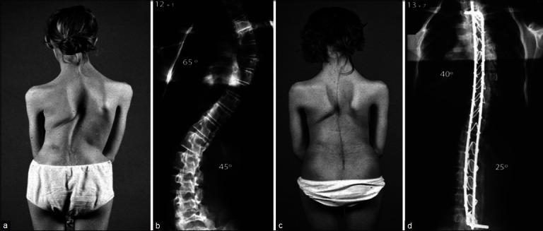
Figure 2.

Clinical photograph (a) and spinal radiograph (b) on a female adolescent patient shows a severe right thoracic and left lumbar scoliosis. (c-d) a posterior spinal fusion with the use of Luque segmental wire/rod instrumentation and autologous iliac crest bone graft produced a balanced spine in the coronal plane with level shoulders and symmetrical waist line
