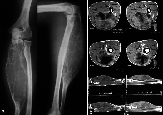
Figure 1.

(a) Preoperative anteroposterior and lateral radiographs showing a well demarcated expansile osteolytic growth involving about two-thirds of ulna. (b) magnetic resonance imaging showing a large circumferential growth from the central medullary canal, expanding the bone. Surrounding cortex is intact and there is no soft tissue component
