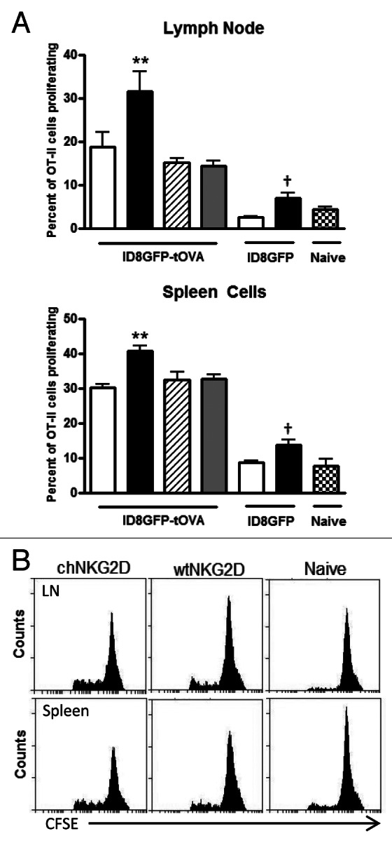
Figure 2. chNKG2D-expressing T cells enhance MHC Class II antigen presentation and the proliferation of CD4+ T cells. (A and B) ID8-GFP or ID8-GFP-tOVA tumor-bearing mice were injected with wtNKG2D-expressing (open), chNKG2D-expressing (filled), interferon γ (IFNγ)-deficient chNKG2D-expressing (hatched), or granulocyte-macrophage colony-stimulating factor (GM-CSF)-deficient chNKG2D-expressing T cells (dotted) i.p. (A) Seven days after T-cell transfer, mediastinal lymph node and spleen cells were isolated and cultured with CFSE-labeled OT-II cells. After 4 d, T-cell proliferation was assessed by flow cytometry. The proliferation of T cells cultured with cells from naïve mice is shown (checked). (B) Representative OT-II CFSE dilution flow cytometry plots as induced by lymph node and spleen cells isolated from naïve mice or from mice bearing ID8-GFP-tOVA tumors treated with wtNKG2D-expressing or chNKG2D-expressing T cells. Data are representative of two individual experiments. The average of each group and SD (n = 8) are shown (**p < 0.01 as compared with ID8-GFP-tOVA tumor-bearing animals receiving wtNKG2D-expressing T cells; †p < 0.05 as compared with ID8-GFP tumor-bearing animals receiving wtNKG2D-expressing T cells).
