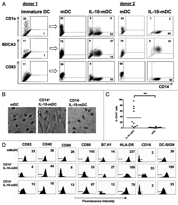
Figure 2. A CD14+BDCA3+ cell population exhibiting macrophage-like morphology and phenotype arises during the maturation of monocyte-derived dendritic cells. (A) Expression of CD1a, CD14, CD83 and BDCA3 on immature monocyte-derived dendritic cells (MoDCs), MoDC matured for 2 d by a mixture of prostaglandin E2 (PGE2), tumor necrosis factor α (TNFα), interleukin (IL)-1β and IL-6 (mDCs), and DCs matured in the presence of IL-10 (IL-10-mDCs). (B) Light microscopic images of mDCs and FACS-sorted CD14+ and CD14− IL-10-mDCs (400× magnification). (C) Percentages of CD14+ DCs matured in the presence (IL-10-mDCs) or absence (mDCs) of IL-10. **p < 0.01 in a Wilcoxon Signed Rank paired analysis. (D) Expression of CD83, CD40, CD80, CD86, B7-H1, HLA-DR, CD16 and DC-SIGN on the indicated mDC subsets. Data are representative of at least eight independent experiments.

An official website of the United States government
Here's how you know
Official websites use .gov
A
.gov website belongs to an official
government organization in the United States.
Secure .gov websites use HTTPS
A lock (
) or https:// means you've safely
connected to the .gov website. Share sensitive
information only on official, secure websites.
