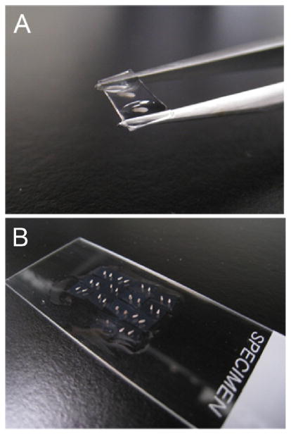
FIGURE 2.
Example of technique used for transferring sections to slides for imaging. (A) Thick sections (150 μm+) can be lifted out of a Petri dish individually, using forceps, without directly touching the embedded tissue. (B) Each sample can then be placed on a slide in a grid fashion for ease of imaging, with or without a coverslip as necessary. When imaging is complete, simply tilt the slide into a scintillation vial and pipette PBT over the surface to wash the samples back into the vial.

