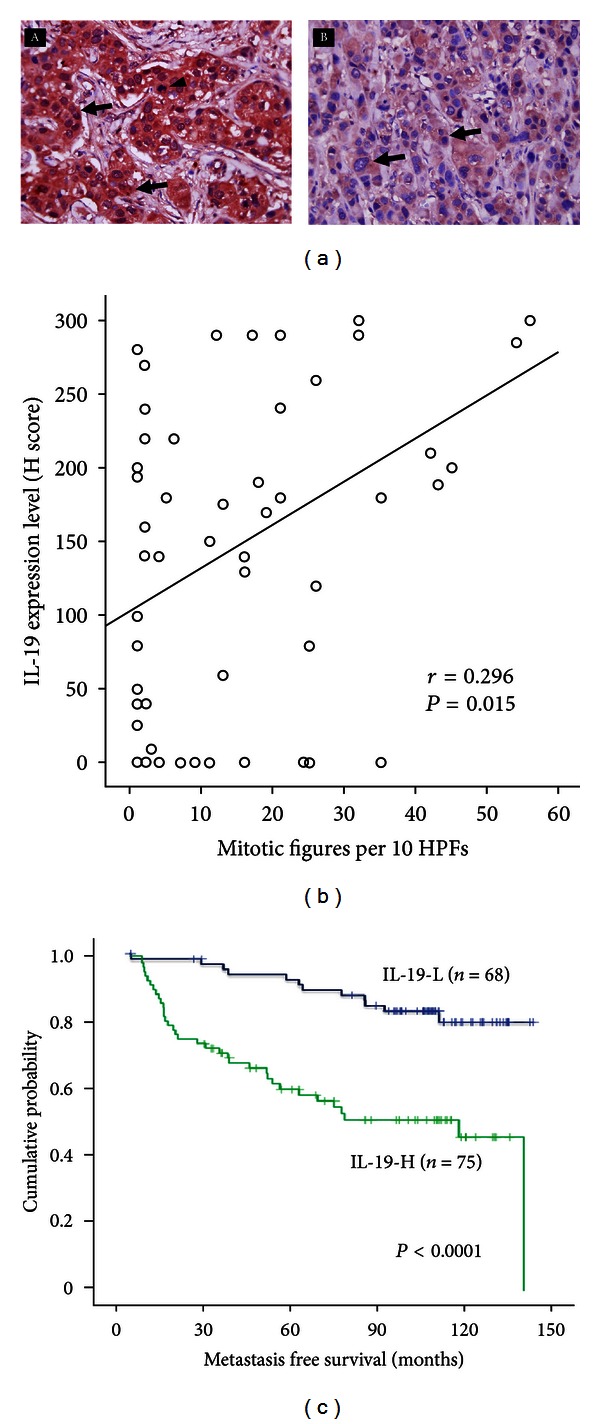
Figure 1.

IL-19 expression in breast tumors was correlated with clinical outcome. (a) Immunohistochemical staining (IHC) showed that IL-19 was strongly (A) or weakly (B) stained in breast invasive duct carcinoma (IDC) cells (arrows) (magnification, ×400). Mitotic figures (A, arrowhead) are commonly found in breast cancer cells strongly stained with IL-19. (b) Mitotic figures were correlated with IL-19 expression levels in breast cancer cells. IL-19 expression levels in 60 IDC tissue samples were analyzed using H scoring. HPFs: high power fields. (c) Of the 143 patients from pathology, Kaplan-Meier plots were used to predict the metastasis-free survival based on IL-19 expression levels. The figure refers to Hsing et al. [13].
