
Fig. 2.
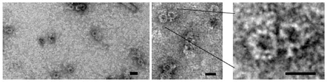
Visualization of ring-shaped VirB complexes. Complexes in peak CsCl fractions 4 and 5 from Fig. 1 were pelleted by ultracentrifugation and resuspended in Lysate buffer (see step 3 in Subheading 3.4). These samples were applied to a carbon-coated grid, stained with 1% uranyl acetate, and analyzed by transmission electron microscopy using a JEOL 1400 microscope. The ring-shaped complexes are similar in size and general architecture to the “core” complexes isolated from the E. coli pKM101 conjugation system by the Waksman laboratory (21, 22). Scale bar: 20 nm.