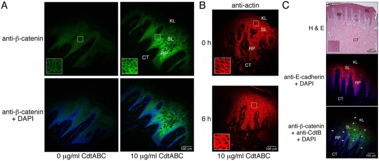
Figure 3.
Localization of cell junction and structural proteins in untreated and CdtABC-treated HGX and gingival tissue from an individual with clinical signs of plaque-induced periodontal disease (PIPD).. (A) Sections from a HGX obtained from a healthy individual were treated with 0 or 10 µg/mL of CdtABC for 18 hrs and then were labeled with a 1:100 dilution of mouse monoclonal anti-β-catenin IgG (Santa Cruz Biotechnology) followed by 5 µg/mL of conjugated goat anti-mouse Alexa Fluor 488 (green fluorescence; Life Technologies). Sections were co-stained with DAPI. Outlined areas are enlarged in the insets. (B) Sections from HGX obtained from a healthy individual were treated with 10 µg/mL of CdtABC for 0 and 6 hrs and were labeled with a 1:100 dilution of rabbit polyclonal anti-actin IgG (Santa Cruz Biotechnology) followed by 5 µg/mL of conjugated goat anti-rabbit IgG Alexa Fluor 594 (Life Technologies). (C) Untreated HGX from an individual with clinical signs of disease. Sections were stained with hematoxylin and eosin and were co-labeled with anti-E-cadherin IgG, anti-β-catenin IgG, anti-CdtB IgG (red fluorescence in bottom panel), and DAPI. Antibody concentrations were the same as those used in Figs. 2 and 3A. The presence of CdtB (arrows) was detected with rabbit polyclonal anti-CdtB IgG (1:50,000 dilution) and 5 µg/mL of conjugated goat anti-mouse Alexa Fluor 488 as described previously (Damek-Poprawa et al., 2011). The explants shown in A and B are from two separate healthy donors. Experiments were performed on healthy and diseased tissue a minimum of 3 and 2 times, respectively, with tissue from different patients.

