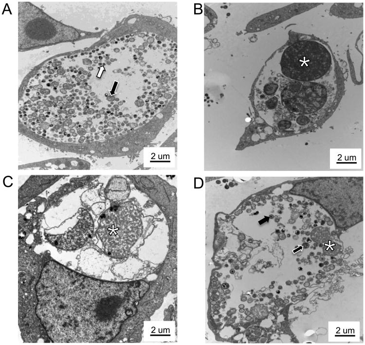
Figure 2.
Amox exposure induces C. muridarum AB formation in culture. Mock or C. muridarum-infected BM1.11 cultures were refed with medium (+/−) amox; 30 h later, some replicates were harvested for TEM while other amox-exposed replicates were refed with either medium or medium (+) amox at 38 hpi and allowed to incubate until 70 hpi. Panel A shows amox (-) infected cells at 38 hpi, panel B shows amox (+) infected cells at 38 hpi, panel C shows amox (+) cells at 70 hpi, and panel D shows cultures exposed to amox from 8 to 38 hpi and then cultured in antibiotic-free medium until 70 hpi. Cell pellets were processed for high contrast TEM as described. Transmission electron micrographs are shown at 7000× magnification; scale bars measure 2 um. RBs are indicated by white-outlined black arrows, EBs by black-outlined white arrows, IBs by solid black arrows and ABs by asterisks. These results are representative of three independent experiments.

