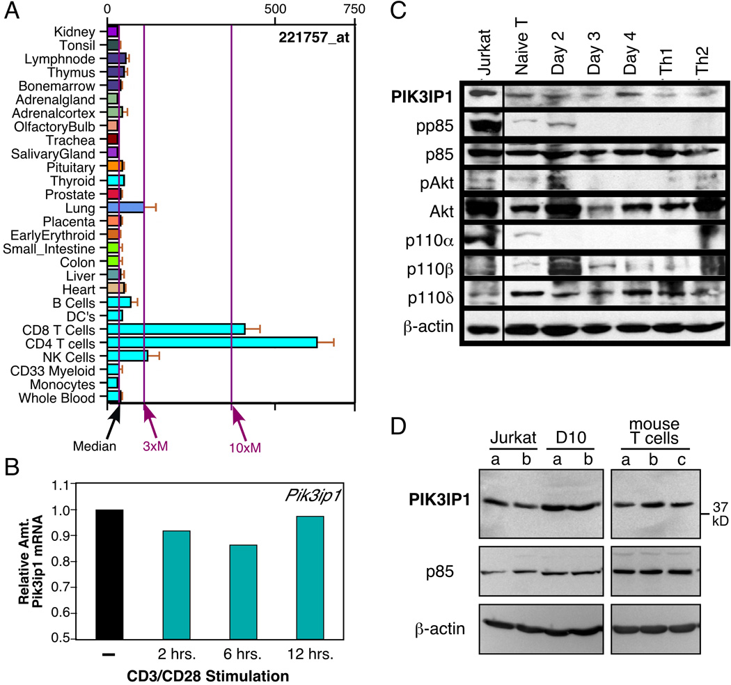
Figure 1.
Expression pattern of PIK3IP1. (A) Analysis of PIK3IP1 message expression in human tissues. Shown are partial results of a BioGPS search for human PIK3IP1. Some tissues have been omitted for clarity, all of which displayed levels of expression at or near the median. (B) Real-time PCR analysis of murine Pik3ip1 message in resting vs. stimulated D10 T cells, a murine Th2 T-cell clone, relative to the amount in resting cells. D10 T cells were stimulated for the indicated times with biotinylated anti-CD3 and anti-CD28 (1 µg/ml each), along with streptavidin (5 µg/ml). Results are representative of three independent experiments. (C) Western blot analysis of expression of PIK3IP1 and other members of the PI3K/Akt pathway in the Jurkat human T-cell line and primary murine T cells, either naïve or activated for the indicated number of days with anti-CD3/CD28 antibodies (1 and 5 µg/ml respectively), under neutral conditions. In addition, bulk cultures of cells stimulated under Th1 or Th2 conditions were also analyzed. PIK3IP1 expression was assessed with a previously described antibody [7]. Blots were also probed with an antibody to beta-actin as a loading control (bottom). Data shown are representative of two independent experiments. (D) Western blot analysis of PIK3IP1 protein in duplicate samples of Jurkat or D10 cells or triplicate samples of primary murine CD3+ T cells, with a commercial antibody (H-180, Santa Cruz Biotech.). p85 and beta-actin were also probed for as controls. Data shown are representative of three independent experiments.

