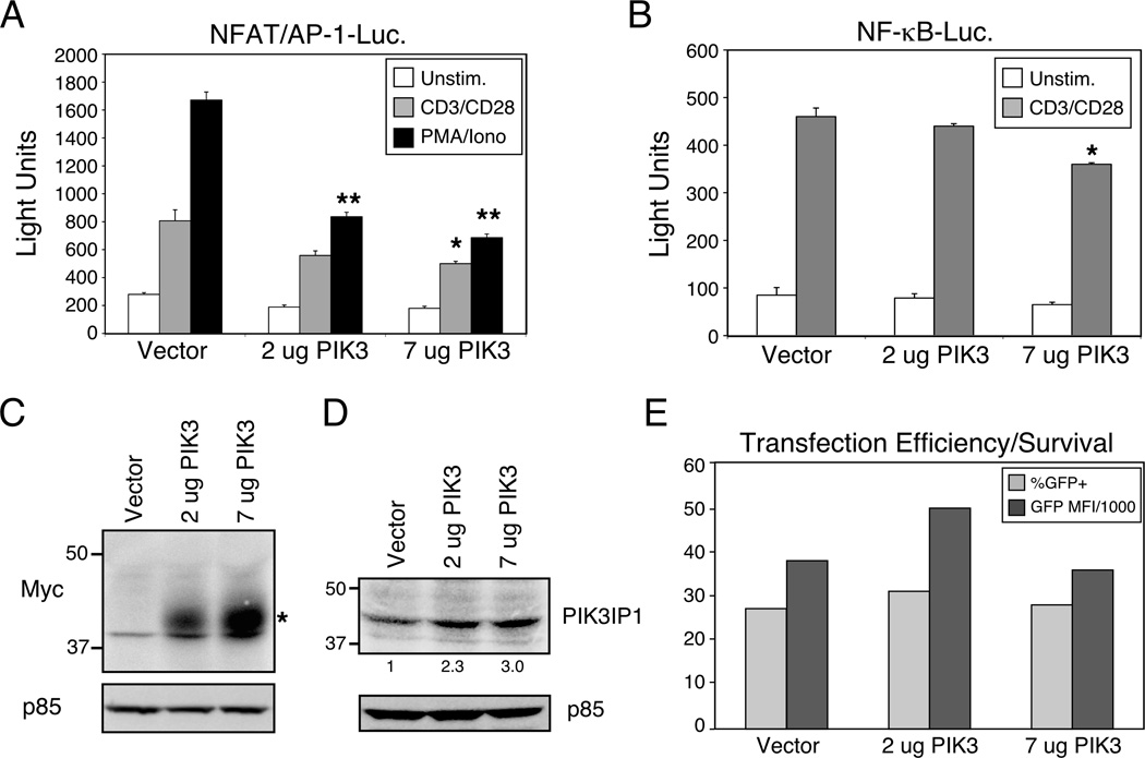
Figure 2.
Suppression of CD3/CD28 signaling by ectopic expression of PIK3IP1. D10 T cells were transfected with either (A) a NFAT/AP-1 or (B) a NF-κB luciferase reporter, a constitutively expressed eGFP construct (as a transfection control) and the indicated plasmids. The next day, cells were stimulated with anti-CD3 and CD28 antibodies (1µg/ml each, plus streptavidin crosslinking (5 µg/ml) or with PMA (50ng/ml) plus ionomycin (1 μM) for six hours, followed by determination of luciferase activity. Data are shown as mean + SD of three replicates from a single experiment, representative of five (NFAT/AP-1) or three (NF-kB) experiments. Bars marked with * or ** indicate a significantly lower response than control vector (p<0.05 or p<0.01 respectively), as determined with the Student’s t test. (C) Expression of myc-tagged PIK3IP1 was confirmed by western blotting with an anti-myc antibody. (D) The degree of PIK3IP1 overexpression was estimated by blotting with a specific polyclonal antiserum that recognizes both endogenous and transfected PIK3IP1. Values under the PIK3IP1 blot indicate relative amounts of protein, compared with vector-transfected cells. (E) Transfection efficiency and survival of the transfected cells were confirmed by flow cytometric analysis of eGFP expression in the same cells. Results are presented as both the percent positive cells (over un-transfected control) and the percent positive x the mean fluorescence intensity (MFI) of the GFP-expressing cells, divided by 1000.

