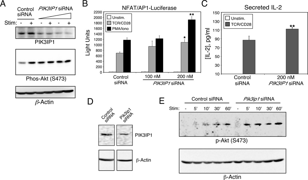
Figure 3.
Enhanced CD3/CD28 signaling by knock-down of PIK3IP1. Jurkat T cells were transfected with an NFAT/AP-1 luciferase reporter and the indicated amounts of PIK3IP1-specific or control siRNA. (A) At 42 hours after transfection, cells were split and either left untreated or stimulated for fifteen minutes with anti-TCR/CD28 antibodies (1 µg/ml and 2 µg/ml respectively). Cells were lysed and the phosphorylation of Akt at serine 473 was then determined by western blotting (middle panel). Cell lysates were also analyzed by western blotting for the expression of PIK3IP1 (upper panel). (B) At 42 hours after transfection, cells were stimulated as indicated for six hours, followed by measurement of luciferase activity. Data are shown as mean + SD of three replicates from a single experiment. Results in panels A and B are representative of three independent experiments. (C) Jurkat T cells were stimulated for 24 hours with anti-TCR/CD28 antibodies, 42 hours after transfection with PIK3IP1 siRNA. Cell-free supernatants were harvested and analyzed by ELISA for production of IL-2. Results are shown as the mean + SD of five experiments. (D–E) D10 T cells were transfected with 200 nM Pik3ip1-specific siRNA, and (D) analyzed 42 hours later by western blotting for PIK3IP1, or (E) stimulated for the indicated times with anti-CD3/CD28 antibodies (1 µg/ml each, plus streptavidin - 5 µg/ml) and analyzed for Akt phosphorylation. Results are representative of two separate experiments. Bars marked with * or ** indicate a significantly higher response than control siRNA (p<0.05 or p<0.01 respectively), as determined with the Student’s t test.

