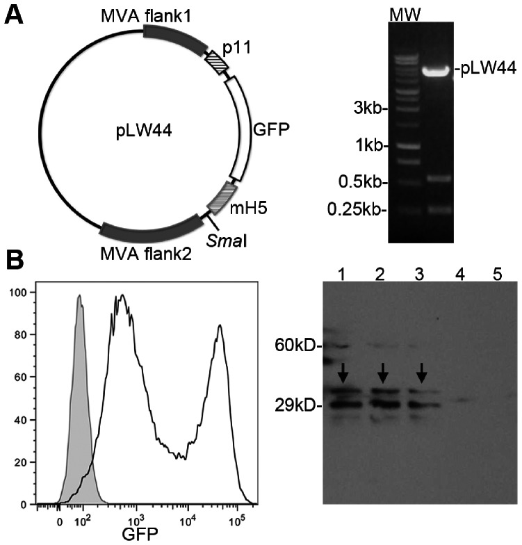
Figure 1. Generation of the recombinant MVA expressing the Surface Antigen 1 (SAG1) of T.gondii.
A, left: plasmid pLW44 was used as shuttle vector for recombination with wild-type MVA genome and insertion of the Sag1 coding sequence into the virus. The Sag1 coding sequence was subcloned in the SmaI site of plasmid pLW44 under control of the promoter mH5 and flanked by two MVA sequences. A, right: digestion of the construct pLW44-SAG1 to verify the correct orientation of the transgene in relation to promoter. B, left: flow cytometry analysis of cells infected with MVASAG1. The presence of recombinant MVA virus is indicated by expression of green fluorescence (FITC channel). In the histogram, gray filled area corresponds to non-infected cells and black line corresponds to MVASAG1 infected cells. B, right: expression of SAG1 in MVASAG1 infected cells. In the Western blot, infected cell lysates were tested against serum from rabbit chronically infected with T. gondii RH strain. Lines 1–3: lysates of cells infected with three different clones of MVASAG1. Line 4: lysate of cells infected with a control MVA expressing only GFP (MVACTRL). Line 5: lysate of non-infected cells. The arrows indicated the bands corresponding to SAG1.

