Fig. 3.
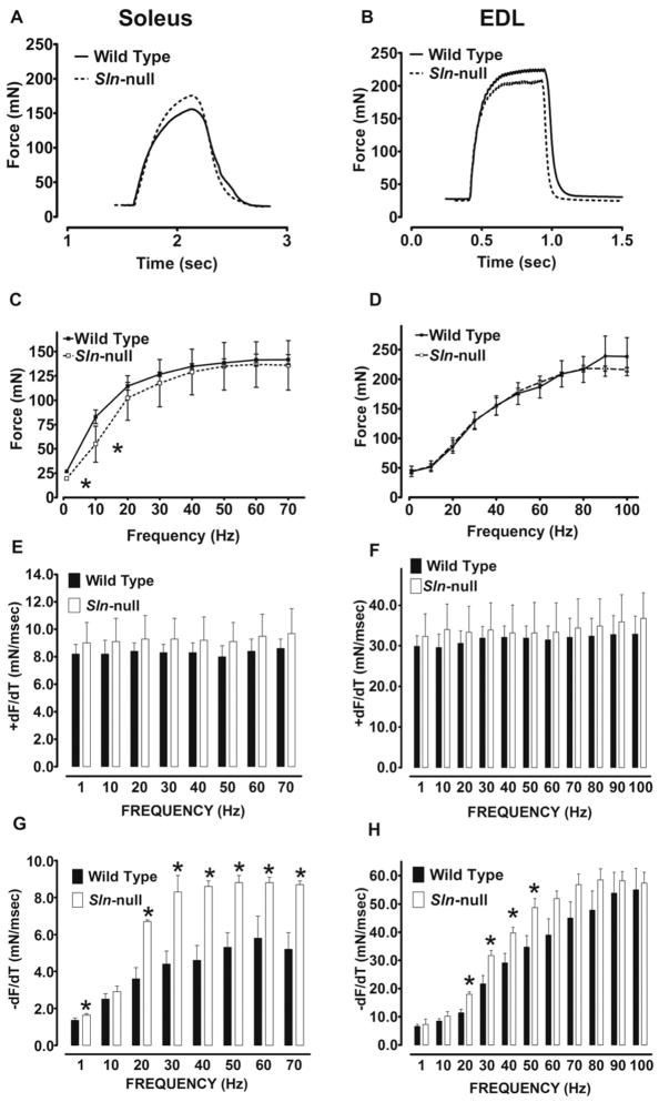
Muscle contractility in soleus and EDL. Isolated skeletal muscles were subjected to force-frequency experiments, and representative tetanic contractions (single 500-ms train at 50 Hz) in soleus (A) and EDL (B) muscles from wild-type and Sln-null mice are shown. The maximum force generated (C and D) and the maximum rates of force development (+dF/dt) (E and F) and relaxation (−dF/dt) (G and H) were determined at each stimulation frequency in soleus (C, E, and G) and EDL (D, F, and H) muscles from wild-type and Sln-null mice. *Significant, P < 0.05 vs. wild type.

