Figure 3.
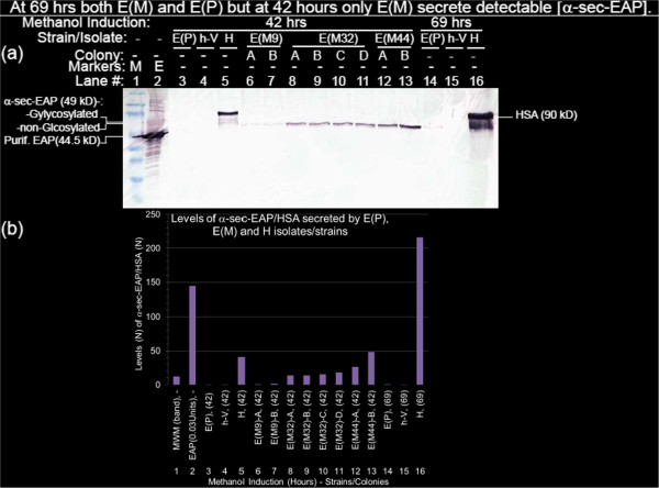
At 69 hrs both E(M) and E(P) but at 42 hours only E(M) secrete detectable [α-sec-EAP]. Culture supernatants of, E(P), h-V and H were harvested after methanol induction for 42 hrs (Figure 3a, lanes 3, 4 and 5) and 69 hours (Figure 3a, lanes 14, 15 and 16), 2–4 colonies per candidate E(M) mutants #9, #32 and #44 (from the re-streaked tertiary (3°) screen e.g. in Figure 2b3) after 42 hours of methanol induction (lanes 6 to 13) were electrophoresed, proteins transferred to NC membranes, sequentially annealed with primary (1°)anti-HSA/primary (1°)anti-EAP) and secondary (2°) anti-Rb-IgG-AP antibodies, washed, reacted with NBT/BCIP substrate and AP terminator as described in the materials and methods section. (Figure 3b). Levels of α-sec-EAP/HSA secreted by E(P), E(M) and H isolates/strains. The filter was scanned and saved as a TIFF file. Pixel densities of the proteins in the regions of interest (ROI) corresponding to, EAP (0.03 U) and MWM standards (Figure 3b, lanes 1, 2), α-sec-EAP (Figure 3b, lanes 3, 4, 6–15) and HSA (Figure 3b, lanes 5 and 16) in the Western Blot shown in Figure 3(a) were quantified with the NIH Image J Densitometric software. The respective pixel densities were normalized to E(P) at 69 Hrs (Figure 3b, lane 14) and plotted with Excel software. Based on this quantification the levels of secretion of α-sec-EAP confirmed the 3 different classes of E(M) mutants visually identified in the initial colony isolation as E(M9)-A, E(M9)-B (Figure 3b, lanes 6, 7), E(M32)-A, E(M32)-B, E(M32)-C, E(M32)-D (lanes 8–11), E(M44)-A and E(M44)-B (Figure 3b, lanes 12, 13).

