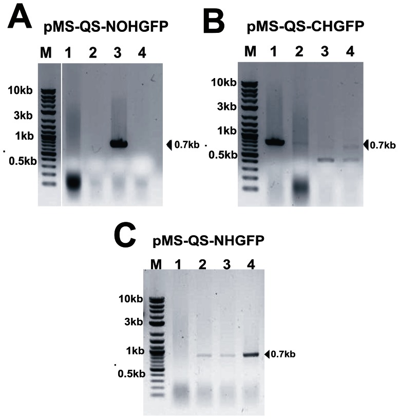
Figure 3. Screening of GFP positive clones using colony PCR.
Agarose gel images after colony PCR showing, (A) clone-3 to be positive in pMS-QS-NOHGFP; (B) clone-1 to be positive in pMS-QS-CHGFP and (C) clones-2, 3 & 4 to be positive in pMS-QS-NHGFP. Vector specific forward primer (PETFor) and gene specific reverse primer (GFPRev-rapid) were used to carry out colony PCR in order to confirm the insertion as well as the orientation of the GFP gene in plasmid.

