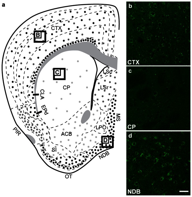
Figure 5. GABARAPL1 expression in the telencephalon.
Immunohistochemical analysis of WT mouse brain tissue with an anti-GABARAPL1 antibody. Images represent typical GABARAPL1 protein expression patterns in neurons found in the cortex-CTX (b), caudate putament-CP (c) and nucleus of the diagonal band-NDB (d). Scale bar represents 20 µm. Abbreviations: ACB (nucleus acumbens), CLA (claustrum), CP (caudate putamen), CTX (cortex), EPd (dorsal endopiriform nucleus), IG (induseumgriseum), LPO (lateral preoptic area), LSr (lateral septal nucleus- rostral part), LSc (lateral septal nucleus- caudal part), MS (medialis strialis), NDB (nucleus of the diagonal band), OT (olfactory tract), PIR (piriform area), SI (substantia innominata).

